

c-Myc (EP121)
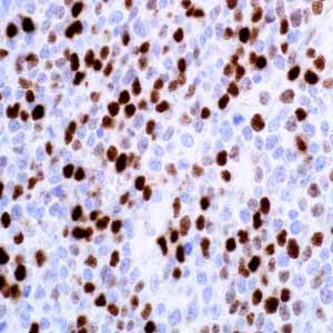
Burkitt lymphoma (BL) can be morphologically indistinguishable from diffuse large B-cell lymphoma (DLBCL).1 The 2008 WHO classification includes a subcategory of aggressive lymphomas characterized by features intermediate between DLBCL and BL in order to accommodate cases in the gray zone between BL and DLBCL. Rearrangement of the MYC gene is found in 3% to 16% of DLBCLs and in nearly 100% of BL.2 Identifying MYC status is important in establishing final diagnosis of DLBCL, BL, or B-cell lymphoma, with features intermediate between DLBCL and BL as well as in differential diagnoses of the lymphomas. Results from one study have shown immunohistochemical (IHC) expression of MYC protein by c-Myc antibody in 219 cases of aggressive DLBCL and 7 cases of BL.1 Nuclear MYC protein expression detected by IHC has shown high sensitivity and specificity in identifying a MYC rearrangement in aggressive DLBCL and BL when at least 70% of lymphoma cells show nuclear positivity.
The IHC c-Myc assay, combined with other antibodies, such as those against CD10, BCL2, and Ki67, is very useful in identifying cases for which MYC FISH analysis is warranted or can be omitted.1 There are reports that nuclear overexpression of MYC protein occur frequently in luminal cells of prostate intraepithelial neoplasia as well as in most primary carcinomas and metastatic disease. However, MYC protein overexpression has yet to be correlated with gain of MYC gene (8q24), suggesting alternative mechanisms for MYC protein overexpression.3