

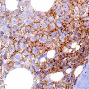
CD38 (SP149)
CD38 molecule is a 46 kDa type II transmembrane glycoprotein with a short N-terminal cytoplasmic tail (20 amino acids) and a long extracellular domain (256 amino acids). CD38 is expressed at low or moderate levels on various hematopoietic cells and in some solid tissues. From a purely morphologic point of view, CD38+ B-lymphocytes are found primarily within the germinal center of secondary lymphoid organs, whereas the marginal zone is predominantly CD38 negative1, although some CD38 weakly +/IgM- cells may be found2. Anti-CD38 behaves as a marker of ontogenesis in T-lymphocytes: ~80% of medullary thymocytes are CD38+, the majority of circulating T-cells are CD38+, and T-cells residing in the tissues are mostly CD38+. Circulating monocytes bear the molecule on their surface, while residential macrophages do not1,2. Functionally active forms of human CD38 are also identified in the outer membrane of red blood cells and on platelets1-3. Among solid tissues, the molecule is expressed by normal prostatic epithelial cells1-3 and by pancreatic islet cells. Other CD38-positive cells include smooth and striated muscle cells, renal tubules, and retinal ganglial cells1-3. Anti-CD38 is extremely useful in classifying functional mature B-lymphocyte subsets. CD38 expression is induced once naive B-lymphocytes are activated, peaks when B-cells enter the germinal center, decreases during centrocyte/centroblast differentiation, and is completely absent in memory B-cells. Therefore, CD38 expression is one of the early markers of mature naive B-cell activation, is upregulated before B-cells enter the germinal center, and undergoes somatic mutations in the IgV genes1-3. From a purely morphologic point of view, CD38+ B-lymphocytes are found primarily within the germinal center of secondary lymphoid organs, whereas the marginal zone is predominantly CD38-1-3. Anti-CD38 is a very useful immunostaining marker, when combined with antibodies against CD138, MUM1, and EMA in a panel, in the diagnosis of immunodeficiency-related lymphomas, which usually include plasmablastic lymphoma, primary effusion lymphoma, and large B-cell lymphoma arising in HHV8-associated multicentric Castleman disease. Such immunodeficiency-related lymphomas are also either pan-B-cellmarker negative or only weakly positive. Furthermore, IHC detection of plasma cells by anti-CD38 immunohistochemical staining on a bone marrow trephine biopsy is necessary to obtain the accurate counts of malignant plasma cells needed to make a definitive diagnosis given that the malignant plasma cell counts are difficult to obtain due to suboptimal bone marrow aspiration, frequent focal distribution of myeloma cells in bone marrow, and loss of neoplastic plasma cells when manual processing is performed. Recent studies have demonstrated that anti-CD38, combined with anti-CD44 (negative) and/or anti-TCL1 (positive), is useful in identifying the cases of large B-cell lymphoma with cMYC gene rearrangement (respective sensitivity of 82% and 77%; respective specificity of 100% and 100%). Therefore, anti-CD38 is very important in differential diagnosis of anti-CD20-positive, anti-TdT/anti-cyclin D-negative diffuse large-to-medium-sized B-cell neoplasms, including diffuse large B-cell lymphoma, Burkitt lymphoma, and B-cell lymphoma, unclassifiable, with features intermediate between DLBCL and Burkitt lymphoma4-7.