

CD44 (MRQ-13)
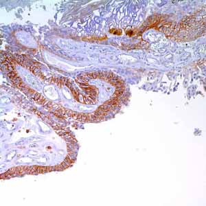
The CD44 family of glycoproteins exists in a number of variant isoforms, the most common being the standard 85-95kD or hematopoietic variant (CD44s) that is found in mesodermal cells such as hematopoietic, fibroblastic, and glial cells, and in some carcinoma cell lines. Higher molecular weight isoforms have been described in epithelial cells (CD44v) and are thought to function in intercellular adhesion and stromal binding. While the other functions and distributions of the CD44 family have not yet been completely elucidated they are also known to participate in embryonic development and angiogenesis as well as other molecular processes associated with specific adhesions, signal transduction, and cell migration. The recent demonstration of a concordance of the cell proliferation nuclear antigen, Ki-67, and CD44 expression in adenomatous polyps, colonic carcinomas and adjacent mucosa raises the possibility of involvement of CD44 in stimulating cell growth. It appears that the CD44-hyaluronate interaction is central to tumor invasiveness; the receptor allowing the uptake and subsequent degradation of metrical hyaluronate. While many human tumors express CD44, a positive correlation between increased CD44v expression and tumor progression and/or dedifferentiation has been demonstrated in only some. Such tumors include non-Hodgkin’s lymphoma (Stauder et al, 1995), hepatocellular carcinoma (Matthew et al, 1996), breast carcinoma, renal cell carcinoma (terpe et al, 1993), colonic carcinoma (Abassi et al, 1993; Wielenga et al, 1993; Herrlich et al, 1995) and some soft tissue tumors (Wand et al, 1996). More recent additions to the list include metastatic melanoma (Sviatoha, et al, 2002), prostatic carcinoma (Ekici et al, 2002), and gastric cancer (Yamaguchi et al, 2002). Conversely, CD44v expression is downgraded in other tumors including neuroblastoma (Shtivelman & Bisho, 1991), squamous cell and basal cell carcinomas of the skin (Herold-Mende et al, 1996). The suggestion that there is a positive association between CD44 isoform expression and progression in human tumors has important implications for diagnosis and prognosis. Unfortunately, the situation is not yet clear-cut. Confusion over the complicated exon boundaries together with the different nomenclature employed by researchers have added to problems of identifying the true metastasis-associated isoforms. Furthermore, stromal cells may contribute to the isoform pattern detected. Probably the most practical application of CD44 immunostaining at present is the discrimination of urothelial transitional call carcinoma-in-situ from non-neoplastic changes in the urothelium (McKenney JK, et al. 2001).